The Human Eye: A Complex Anatomy
The human eye is a single, functional unit that is itself very complex. In order for us to see, proper anatomical development of the eye is very important, because any defects in the eye can drastically affect our vision.
As light enters the eye, it first passes through a clear, transparent lens called the cornea. The cornea, much like a camera or eyeglass lens, is curved, allowing it to focus the light as it enters the eye. In fact, the cornea is the major refractive element of the eye, meaning it brings most of what we see into focus.
The light then passes through the hole in the iris, called the pupil, where it is further focused by the eye’s lens.
At the very back of the eye is the retina, composed of tiny, cellular rods and cones, which allow us to sense light and color. When light hits the retina, the rods and cones produce electrical impulses that are carried to the brain via the optic nerve, and ultimately the visual pathway.
If it seems like that’s a lot of information to take in, that’s because it is. Luckily, the eye can be divided into smaller external and internal parts to help make it simpler to understand.
External Ocular Anatomy
(This list is organized from outermost to innermost parts of the eye.)
Conjunctiva
The conjunctiva is a continuous, transparent mucous membrane, which lines the inner surface of the eyelids (and the outer surface of the eyes) to allow the eyelids to open and close.
EXTRAOCULAR MUSCLES
This is a group of 7 striated (fibrous, banded) muscles that connect to the sclera (see below) to control the movement of the eyes with rapid precision.
The 6 muscles that control the eye are:
- Medial
- Lateral
- Superior rectus
- Inferior rectus
- Superior oblique
- Inferior oblique
The 1 muscle that controls the eyelids is the: Levator palpebrae superioris.

Each of these muscles is in turn connected to one of 3 cranial nerves—the Oculomotor nerve (3rd), the Trochlear nerve (4th), or the Abducens nerve (6th)—which allow the eyes to move.
Sclera
The sclera is commonly called the “white of the eyes.” It’s the tough outer layer of the eye, made of collagen to help the eye maintain its shape. The sclera covers almost the entire surface of the eye and is where the tendons of the extraocular muscles (see below) are attached.
Cornea
The cornea is the clear, dome-shaped anterior (front) part of the eye that is responsible for two-thirds of the refractive power of the eye. It’s made up of collagen fibers that are cross-linked at such a microscopic level that the cornea appears to be transparent. This allows light to enter the eye so that it is focused on the retina (see below).

Internal Ocular Anatomy
(This list is organized from anterior (front) to posterior (back) of the eye.)
Anterior Chamber
The anterior chamber is the space between the cornea and the iris, which is filled with a fluid called aqueous humor.
AQUEOUS HUMOR
Aqueous humor is a clear, watery liquid (like plasma), created by the ciliary body. This is what helps the eye maintain a consistent spherical shape. The aqueous humor also provides nutrition to the cornea in the form of sugars, vitamins, proteins, etc.
IRIS
The iris is the uniquely colored part of the eye. It is a smooth, disc-shaped muscle with an opening (the pupil) in the center. The iris is used to dilate the pupils (make them wider or narrower) according to the light around you. As the iris causes the pupils to dilate, they let in more or less light to the retina in the posterior (back) part of the eye.
PUPIL
The pupil is the opening in the center of the iris, which allows light to enter the interior of the eye.
LENS
The lens of the eye is located directly behind the pupil. The lens is responsible for the remaining one-third of refractive power of the eye, once the light passes through the cornea (see above). The lens changes its shape to focus light onto the retina and aids in focal differentiation of near objects.
CILIARY BODY
The ciliary body is attached to the outer edge of the iris. It produces the aqueous humor (see above) and helps to change the shape of the lens when focusing.
VITREOUS
The vitreous is a cavity that lies between the lens and the retina and makes up the majority of the space inside the posterior (back) of the eye. The vitreous is filled with a gelatinous substance known as the vitreous humor, which nourishes the innermost structures of the eye. Light comes into the eye through the pupil, passes through the vitreous, and is projected onto the retina.
RETINA
The retina is a thin, transparent structure that covers the inner wall of the posterior (back) eye. Light enters the eye and is focused onto the retina, where the image is converted into a chemical signal by microscopic rods and cones, before being transmitted through the optic nerve to the brain. The retina is a very complex structure with layers of different specialized cells.
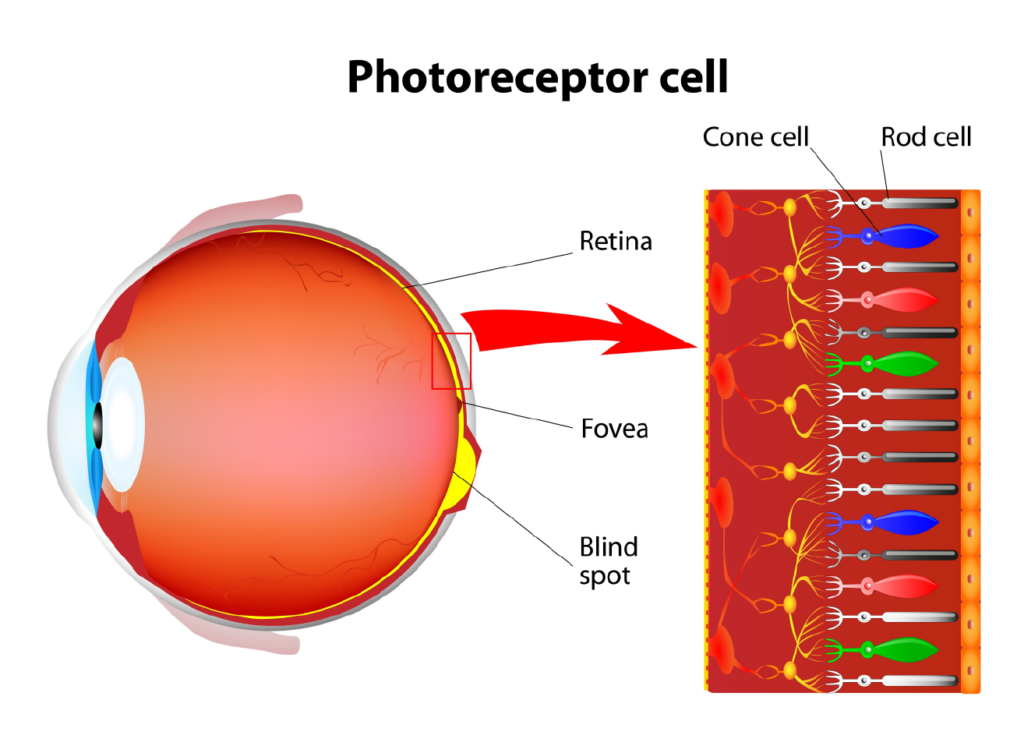
PHOTORECEPTORS
Photoreceptors are a group of highly-specialized cells in the retina that receive light and convert it into chemical impulses that can be transmitted via nerve cells to the brain. There are 2 different types of photoreceptors: rods and cones. Rods perceive the contrast between black and white and are used primarily for night vision. Cones are specialized in red, blue, and green colors and are responsible for our color perception and central vision.